• Skip to main content
  • Skip to header right navigation
  • Skip to site footer

786-300-3183 | [email protected]

  • Twitter
  • Facebook
Florida Society of Anesthesiologists

Florida Society of Anesthesiologists

  • About FSA
    • FSA Leadership
      • FSA Past Presidents
      • Distinguished Service Award Past Recipients
      • Recipients of the FSA Presidential Engagement Award
    • FSA Staff
    • FSA NEWS
    • Calendar of Events
    • Contact FSA
    • FSA Charter & Bylaws
    • FSA Speakers Bureau
  • FSA Annual Meeting
    • 2025 Annual Meeting
    • 2024 Annual Meeting Recap
    • Call For Abstracts
    • Past Posters
      • 2025 FSA Podium and Poster Abstracts
      • 2024 FSA Podium and Poster Abstracts
      • 2023 FSA Podium and Poster Abstracts
      • 2022 FSA Podium and Poster Abstracts
      • 2021 FSA Posters
      • 2020 FSA Posters
      • 2019 FSA Posters
      • 2018 FSA Posters
    • Past Meetings
      • 2023 Meeting Recap
      • 2022 Annual Meeting Recap
      • 2019 Annual Meeting Recap
      • 2018 Annual Meeting Recap
  • FSAPAC
    • Donate to the FSAPAC
  • Member Login
  • Member Portal
  • Become a Member
    • FSA Membership Renewal
    • Join the Florida Society of Anesthesiologists (FSA)

2018 FSA Posters

2018 FSA Posters

P057: AORTIC ROOT ABSCESS AND THE LOST ART OF THE PHYSICAL EXAM
Andres Bacigalupo Landa, MD, Jayanand D'Mello, MD; Mount Sinai Medical Center

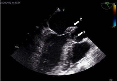
A 69-year-old female patient was admitted to the hospital with malaise and abdominal pain. On hospital day five, the patient developed signs of sepsis and new-onset atrial fibrillation along with a quickly progressing cardiac conduction abnormality. Repeat imaging revealed acute cholecystitis, and emergency surgery was scheduled. In the holding area, the anesthesiologist performed a cardiac physical exam, which revealed a previously undocumented diastolic murmur. This new finding prompted the use of an intraoperative transesophageal echocardiogram (TEE), which subsequently revealed an aortic root abscess with an aortic valve vegetation (figure 1) and severe aortic insufficiency. Twelve days later, the patient underwent an aortic valve replacement. This case underscores the importance of physical exam of the anesthesiologist as it may reveal imperative information that may have been previously missed by other clinicians and highlights the importance of the utilization of the intraoperative TEE as part of the anesthesiologist’s skill set.

Intraoperative TEE (mid-esophageal long axis view of the AV) showing an AV vegetation (arrow 1) and an AV abscess (arrow 2).

Copyright © 2025 · Florida Society of Anesthesiologists · All Rights Reserved